![]()
![]()
![]()
![]()
Contact: Dr. rer. nat. André Haase
Our research group focuses on the translational tumor biology of retinoblastoma and colorectal carcinoma. Our overarching goal is to elucidate molecular mechanisms of tumor progression, identify novel therapeutic targets, and develop innovative treatment strategies that ultimately contribute to improved patient care.
Two Tumor Entities – A Complementary Research Strategy
Retinoblastoma is a rare, highly aggressive pediatric ocular tumor characterized by an exceptionally homogeneous genetic background. This unique feature makes retinoblastoma an ideal model system to investigate fundamental mechanisms of tumor initiation, metabolic reprogramming, and therapy resistance under well-defined genetic conditions.
In contrast, colorectal carcinoma represents one of the most genetically heterogeneous malignancies and is among the most common cancers worldwide. The pronounced molecular diversity of these tumors reflects the clinical reality of personalized oncology and allows us to evaluate novel therapeutic approaches under highly variable and clinically relevant conditions.
The parallel investigation of both tumor entities enables us to translate fundamental oncogenic principles identified in a genetically well-defined model system (retinoblastoma) into a highly complex clinical scenario (colorectal carcinoma). This strategy allows us to identify robust, tumor-independent mechanisms while simultaneously developing individualized therapeutic approaches.
Our Research Projects
A particular emphasis is placed on the functional validation of newly identified targets and the preclinical assessment of novel compounds.
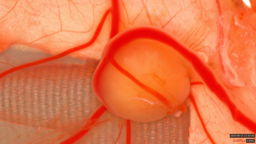
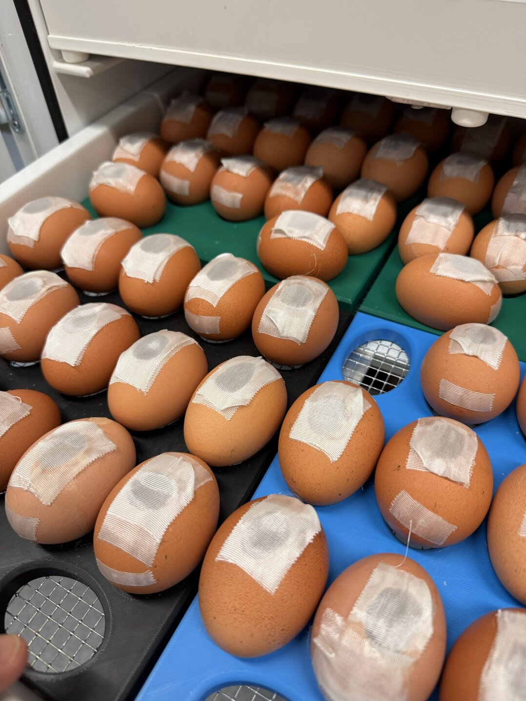
Model System: The Chorioallantoic Membrane of the Chicken Embryo
The chorioallantoic membrane (CAM) is a thin, highly vascularized membrane surrounding the chicken embryo and physiologically functions as a “lung-like” organ. It enables gas exchange and is therefore strongly perfused and metabolically highly active.
These unique biological properties make the CAM an ideal in ovo model system for tumor research. Human tumor cells or tumor tissues can be directly grafted onto the membrane, where they rapidly engraft, become vascularized, and form solid tumors within a few days.
The CAM model allows the investigation of tumor growth, neovascularization (angiogenesis), invasion, and metastasis in a living organism. At the same time, it enables preclinical testing of novel compounds and therapeutic strategies under physiologically relevant conditions.
Due to its short experimental duration, high reproducibility, and application prior to the pain-sensitive developmental stage of the embryo, the CAM model meets the ethical requirements of the 3R principles to an exceptional degree and represents a bridge between classical cell culture systems and conventional animal models.
Within the CAM model, we are able to:
By doing so, we close the gap between traditional cell culture models and labor-intensive animal models and establish a powerful platform for translational cancer research.
Our research group integrates state-of-the-art cell and molecular biology with innovative tumor models, functional imaging, and pharmacological development. Through the close interplay of basic research and preclinical validation, we create a strong foundation for future personalized therapeutic strategies in oncology.
Copyright © pharmacology 2026
Last update: Feb 02, 2026

